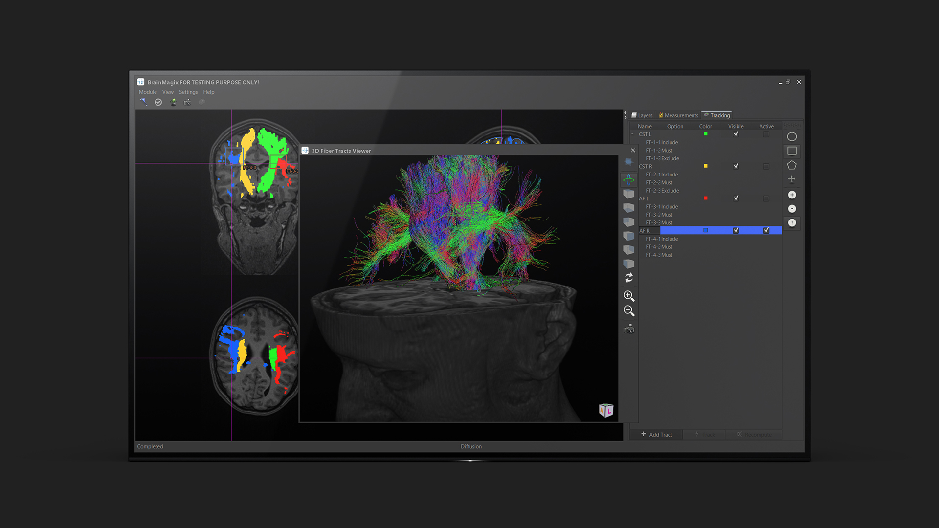
Tractography
Issue of Crossing Fibers
However, the most widespread implementation of the technique, based on the diffusion tensor imaging (DTI) model, has limitations (Farquharson et al., 2013). The model assumes that, in each voxel, there is a unique orientation of the fibers, the direction of which is represented by the tensor’s main eigenvector (Mori and Tournier, 2013).
This assumption is not valid in case of crossing fibers. The term “crossing fibers” generally refers to regions in which the fibers’ orientation is not unique, i.e. when the fibers are interdigitating, brushing past each other, curving, bending or diverging (Mori and Tournier, 2013). Given the relatively large voxel size on diffusion images, the proportion of white matter voxels in the brain that contain multiple fiber orientation has been reported to reach up to 90% (Jeurissen et al., 2013).
Tracking of the Pyramidal Tract
Even the pyramidal tract (i.e. the corticospinal (CST) and corticobulbar tracts), which is the mostly widely tracked in the context of neurosurgical planning, can only be partially identified with the DTI-based method (Farquharson et al., 2013; Lee et al., 2015; Mormina et al., 2015). Its lateral portion, corresponding to the somatotopic representation of hand, face, tongue, and voluntary swallow muscles, is usually not detectable by DTI-based tractrography (Mormina et al., 2015). This is partially due to the crossing fibers that DTI cannot resolve, at the level of its intersection with the superior longitudinal fasciculus (SLF) (Lee et al., 2015). Therefore, relying on DTI tractography for surgery planning has been shown in some cases to lead to entirely avoidable loss of motor function (Kinoshita et al., 2005).
Constrained Spherical Deconvolution
The method of spherical deconvolution (Tournier et al., 2004) can be used to estimate the distribution of fiber orientations present within each imaging voxel. With this method, the signal measured during a high angular resolution diffusion weighted imaging (HARDI) session can be expressed as the convolution, over spherical coordinates, of the response function (RF), representing the signal of a single coherently oriented population of fibers, with the fiber orientation distribution (FOD). The FOD can be then obtained by the inverse operation, i.e. the spherical deconvolution of the diffusion weighted signal by the response function. Deconvolution methods are very sensitive to noise effects, but the robustness of the operation can be greatly enhanced by a non-negativity constraint (Tournier et al., 2007), (hence the name: constrained spherical deconvolution), leading to a robust determination of the fiber orientations within a clinically acceptable time (typically under 10 minutes) (Farquharson et al., 2013; Mori and Tournier, 2013). Several studies have demonstrated the superiority of the method, when compared with DTI-based tractrography, in the context of neurosurgical planning (Farquharson et al., 2013; Mormina et al., 2015).
References
Farquharson et al. White matter fiber tractography: why we need to move beyond DTI. J. Neurosurg. 2013, 118: 1367–1377.
Golby et al. Interactive diffusion tensor tractography visualization for neurosurgical planning. Neurosurgery 2011, 68: 496–505.
Jeurissen et al. Investigating the prevalence of complex fiber configurations in white matter tissue with diffusion magnetic resonance imaging. Hum. Brain Mapp. 2013, 34: 2747–2766.
Kinoshita et al. Fiber-tracking does not accurately estimate size of fiber bundle in pathological condition: initial neurosurgical experience using neuronavigation and subcortical white matter stimulation. NeuroImage 2005, 25: 424–429.
Lee et al. Have You Ever Seen the Impact of Crossing Fiber in DTI?: Demonstration of the Corticospinal Tract Pathway. PLoS ONE 2015, 10.
Mori and Tournier Introduction to Diffusion Tensor Imaging: And Higher Order Models (Academic Press).
Mormina et al. MRI Tractography of Corticospinal Tract and Arcuate Fasciculus in High-Grade Gliomas Performed by Constrained Spherical Deconvolution: Qualitative and Quantitative Analysis. AJNR Am. J. Neuroradiol. 2015, 36: 1853–1858.
Tournier et al. Direct estimation of the fiber orientation density function from diffusion-weighted MRI data using spherical deconvolution. NeuroImage 2004, 23: 1176–1185.
Tournier et al. Robust determination of the fibre orientation distribution in diffusion MRI: Non-negativity constrained super-resolved spherical deconvolution. NeuroImage 2007, 35: 1459–1472.