Perfusion Magnetic Resonance Imaging
Contrast Agent
Perfusion MRI is based on the analysis of the contrast enhancement of MRI images after a peripheral injection of a contrast agent, e.g. Gd-DTPA. The injection of 0.1 mmol/kg is usually quick (i.e. a bolus), at a rate of 5-10 ml/s, and is followed by a saline flush.
Dynamic Susceptibility Contrast
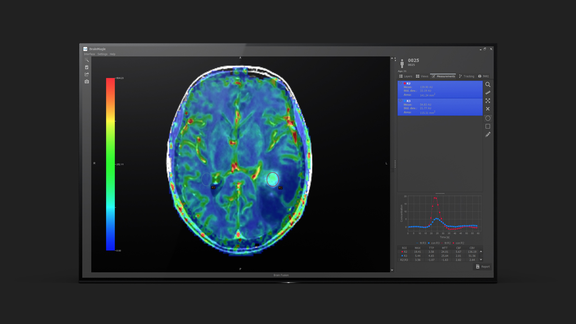
Dynamic Susceptibility Contrast (DSC) perfusion MRI uses a GRE-EPI (T2*-weighted) sequence in which the contrast agent induces an hypointensity. For example, 40 volumes can be acquired every 1.5 seconds during 1 minute. Concentration-versus-time curves are calculated from the variation of the MR image signal induced by the contrast agent. The tissue curves are sometimes deconvolved by the arterial input function (AIF) in order to eliminate the influence of the injection kinetics. From the curves, several parameters can be calculated: cerebral blood volume (CBV), cerebral blood flow (CBF), mean transit time (MTT), time to peak (TTP), time to peak of the residue function (Tmax), etc.
Dynamic Contrast Enhanced
Dynamic Contrast Enhanced (DCE) perfusion MRI uses a spoiled fast gradient echo (T1-weighted) sequence in which the contrast agent induces an hyperintensity. Conversion from image signal to concentration-versus-time curves requires a calibration procedure, e.g. a measurement of pre-injection T1 relaxation time by relaxometry. Taking into account the arterial input function (AIF), tracer kinetics models applied to the curves lead to a measurement of the transfer constant between the intra-vascular and extra-vascular spaces Ktrans (a marker of vessel permeability) and of the fractional volume of the extra-vascular extra-cellular space.
Applications
DSC perfusion MRI is often used in combination with diffusion MRI for the imaging of ischemic stroke. The mismatch between perfusion and diffusion MRI is a marker of the ischemic penumbra (i.e. the zone which can still be preserved by an appropriate treatment).
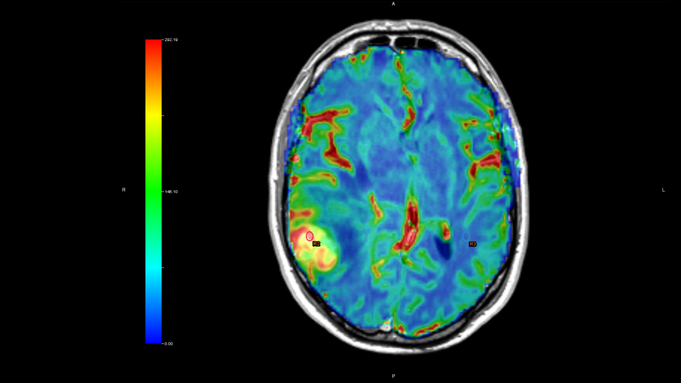
In brain tumors, DSC and DCE perfusion can be used to assess the tumoral vascularization, as a marker of the tumor's grade.
References
Essig et al. Perfusion MRI: the five most frequently asked technical questions. AJR Am J Roentgenol. 2013, 200:24-34.